Neuroendocrine adenoma of the middle ear (NAME) is a rare neoplasm of the middle ear, comprising less than 2% of all middle ear tumors. The clinical presentation of NAME is often nonspecific and can include unilateral hearing loss, aural fullness, tinnitus, vertigo, and facial palsy. Diagnosis of NAME can be challenging, as a pre operative clinical diagnosis is difficult to make. Definitive diagnosis is based on histopathology and immunohistochemistry (IHC) findings. In this case report, we present the case of a 41-year-old woman who presented with a one-year history of unilateral hearing loss and tinnitus on the right side. Examination with an otoscope revealed a pale-reddish mass in the right external auditory canal. Pure tone audiometry showed right conductive hearing loss with a 12.5 dB air-bone gap. Computed tomography revealed a soft tissue lesion in the right external auditory canal, middle ear, attic space, and aditus ad antrum, encasing of ossicles. Magnetic resonance image
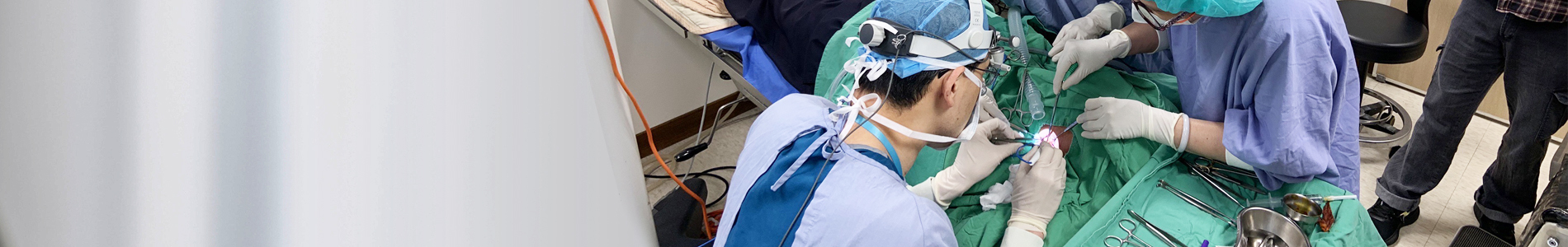
displayed a tumor with isointense to hyperintense signal in T1 and T2-weighted image with contrast enhancement. The initial diagnostic impression was paraganglioma, and surgical intervention was deemed necessary. The tumor was excised through a post auricular approach with the assistance of a microscope and endoscope, revealing that the tumor was occupying the middle ear, attic space, and aditus ad antrum. A rigid endoscope was used to confirm the absence of residual tumor. The result from an intraoperative frozen section favored a paraganglioma. However, a postoperative histological and immunohistological examination confirmed the tumor as a NAME. The post-operative air-bone gap decreased to 5 dB. The patient underwent regular follow-up for 2 years, during which no evidence of recurrent disease was observed. We conclude that endoscope-assisted microscopic surgery is effective in reducing residual lesions and achieving complete tumor resection while preserving the ossicles. Given that NAME is considered a low-grade malignancy, long-term follow-up is necessary. (J Taiwan Otolaryngol Head Neck Surg 2023; 58:143-149)